Global healthcare systems are facing an expanding disparity between the demand for diagnostics and the availability of specialists. This is particularly visible in neuroradiology, where the volume of brain MRIs is increasing, but shortages of radiologists, reporting backlogs, and errors due to weariness jeopardize patient safety. In this context, the ALIGN Study by Wood et al. signifies a crucial advancement in the development of artificial intelligence (AI) for medical imaging.
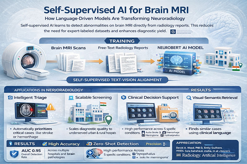
The paper, published in Radiology: Artificial Intelligence, presents a self-supervised text-vision alignment system for the automatic detection of brain MRI anomalies, trained directly on regular scans and unannotated neuroradiology reports, eliminating the necessity for expert-labelled picture datasets. This methodological transition is not incremental; it is fundamental.
Transcending the limitations of labelled data
Traditional medical AI has predominantly depended on supervised learning, necessitating radiologists to manually annotate thousands of images. This method is costly, sluggish, and progressively indefensible within overburdened healthcare systems. The ALIGN Study tackles this impediment by utilizing a plentiful although underexploited resource: radiological reports. These reports have comprehensive semantic descriptions created by specialists and are intrinsically associated with imaging data in hospital records.
By correlating MRI images with the semantic embeddings of reports through a specialized neuroradiology language model (NeuroBERT), the framework discerns the characteristics of “normal,” “abnormal,” and disease-specific patterns—absent specified categories. This enables the system to adjust to novel clinical inquiries without the necessity of retraining on newly annotated datasets.
The findings are persuasive. Wood et al. present an AUC of 0.95 for the classification of normal versus abnormal cases, demonstrating strong generalization across four external institutions, notwithstanding variations in scanners, protocols, and patient demographics. The zero-shot performance concerning clinically significant entities, including acute stroke, cerebral haemorrhage, multiple sclerosis, meningioma, and hydrocephalus, further highlights the adaptability of this methodology. These findings combined indicate that self-supervised AI has transitioned from a theoretical concept to a therapeutically applicable paradigm.
Clinical significance: triage, safety, and operational resilience
The immediate healthcare benefit of such a system resides in intelligent triage. In practical radiology, the urgency of scans varies significantly. An AI system that accurately differentiates between normal and abnormal tests can prioritize time-sensitive situations, thereby minimizing delays in the management of strokes or haemorrhages.
The work also effectively illustrates visual-semantic image retrieval, allowing doctors to access analogous previous cases through normal clinical language. This affects not just diagnostic assistance but also education and quality assurance, enabling radiologists and trainees to contextualize findings within institutional experience.
In nations such as India, where the ratio of radiologists to the population is significantly low and advanced neuroradiology expertise is inequitably allocated, scalable AI techniques could have a transformative impact. Self-supervised frameworks reduce reliance on human annotation and the availability of subspecialists, so facilitating more equitable access to diagnostic quality without sacrificing professional monitoring.
An equitable and accountable contribution
The authors are judicious in their conclusions. They explicitly recognize limits, such as the present autonomy of sequence-specific models and the necessity for future multisequence reasoning. This transparency enhances the credibility of the work and conforms to the standards of responsible AI deployment.
The ALIGN Study distinctly categorizes AI as a tool for clinical decision assistance rather than as a means for autonomous diagnosis. This differentiation is crucial. The genuine potential of AI in healthcare resides in augmentation—alleviating cognitive burden, enhancing prioritization, and fostering consistency—while maintaining human knowledge at the core.
Gratitude towards the authors and the journal
The authors of the ALIGN Study—Wood et al.—merit genuine commendation for providing a methodologically sound, therapeutically relevant, and progressive contribution. The dataset’s magnitude, incorporation of potential external validation, and emphasis on practical applicability demonstrate a profound comprehension of both technical and clinical contexts. The study addresses a significant obstacle in medical AI—data labelling—thereby paving the way for sustained innovation in radiography.
Equal recognition is warranted for Radiology: Artificial Intelligence for its role in selecting and disseminating such significant work. The journal consistently establishes a standard by emphasizing research that is both technically advanced and ethically sound, as well as clinically significant. This solidifies its leadership in determining the integration of AI into routine medical practice.
Final Analysis
The ALIGN Study is a significant turning point in neuroradiology artificial intelligence. Wood et al. have illustrated a feasible approach beyond fragile, label-dependent models by proving that machines can learn directly from clinical language at scale. This work provides not only innovation but also guidance for healthcare systems facing increasing diagnostic demands, steering them towards flexible, scalable AI that corresponds with the realities of patient care.
Dr. Prahlada N.B
MBBS (JJMMC), MS (PGIMER, Chandigarh).
MBA in Healthcare & Hospital Management (BITS, Pilani),
Postgraduate Certificate in Technology Leadership and Innovation (MIT, USA)
Executive Programme in Strategic Management (IIM, Lucknow)
Senior Management Programme in Healthcare Management (IIM, Kozhikode)
Advanced Certificate in AI for Digital Health and Imaging Program (IISc, Bengaluru).
Senior Professor and former Head,
Department of ENT-Head & Neck Surgery, Skull Base Surgery, Cochlear Implant Surgery.
Basaveshwara Medical College & Hospital, Chitradurga, Karnataka, India.
My Vision: I don’t want to be a genius. I want to be a person with a bundle of experience.
My Mission: Help others achieve their life’s objectives in my presence or absence!
My Values: Creating value for others.
Reference:
Leave a reply


Leave a reply